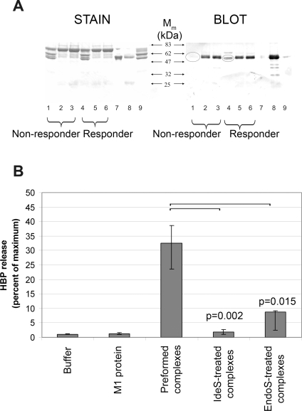
Figure 5. M1 protein forms HBP-releasing precipitates with fibrinogen and IgG in responder plasma.
(A) Purified M1 protein was added (10 µg/ml) to the plasma of a responder or a non-responder (diluted 1∶10). After incubation, the resulting precipitates were spun down and the supernatants were saved. Precipitates were washed, boiled in sample buffer and subjected to SDS-PAGE (lanes 1 and 4). The supernatants (see above) were run in lanes 2 and 5, human plasma diluted 1∶500 was run in lanes 3 and 6, M1 protein (1.2 µg) in lane 7, human polyclonal IgG (2 µg) in lane 8, and human fibrinogen (2.6 µg) in lane 9. One gel was stained with Commassie Blue (STAIN), whereas an identical gel was blotted onto an Imobilon filter and probed with goat antibodies against human IgG (BLOT). (B) Whole blood diluted 1∶10 from a non-responder was used. Buffer or M1 protein (1 µg/ml) was added. M1 protein was pre-incubated with responder plasma and the complexes formed were also tested. Such complexes were also digested with IdeS or EndoS and subsequently added to the blood. The release of HBP was measured and the data shown represent the median±interquartile range from six separate experiments. The HBP release from blood supplemented with complexes was compared to the IdeS- or the EndoS- treated complexes using the Mann-Whitney U test, and the result is shown as p values uncorrected for multiple comparisons.

