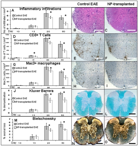
Figure 5. Attenuation of the progression of inflammation and tissue damage in the CNS of NPs-transplanted mice.
Pathological examination of spinal cord sections from NP-transplanted and control mice were performed at 10, 13, 20 and 50 days post EAE induction to evaluate CNS inflammation, demyelination and axonal damage. In NP-transplanted mice an attenuation of the number of immune-cell infiltrates (A), T cells (D) and macrophages/activated microglia (G) per mm2 was evident from day 13 post EAE induction and became significant at days 20 and 50. Demyelination and axonal damage, which were analyzed by loss of Kluver Barrera (J), and Bielschowsky staining (M), respectively, were both significantly reduced at day 20 and 50 post EAE induction. The differences between the study and control groups in the severity of all parameters gradually increased with time. Representative day 20 images of H&E staining (B, C), immunostaining for CD3 (E, F) and Mac3 (H, I), Kluver Barrera staining (K, L) and Bielschowsky silver staining (N, O). * P<0.05. Scale bars: 100 µm.

