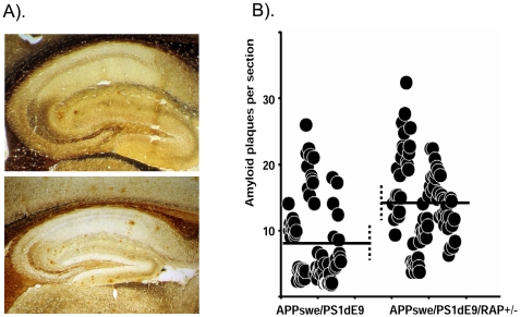
Figure 1. Partially reducing RAP increases Aβ deposition in the brains of APPswe/PS1dE9 mice.
A). Silver staining reveals increased amyloid deposition in the brains of [APPswe/PS1dE9](+/−)/RAP(+/−) mice as compared to the parental APPswe/PS1dE9 line 85 mice. B). Plot of the results from counting amyloid plaques following procedures described in Methods. Each dot indicates the number of amyloid plaques on a section. Six sections per animal were counted by two independent assessors that were blind to the genotype of the animals. Each section was counted 3 times by each assessor and averaged. Statistical analyses was conducted on the mean number of deposits for each animal [n = 6 for APPswe/PS1dE9 mice and n = 7 for the [APPswe/PS1dE9](+/−)/RAP(+/−) mice]. The statistical difference in amyloid burden between animals of the 2 genotypes was estimated by 2-tailed t-test with equal variance (p<0.05).

