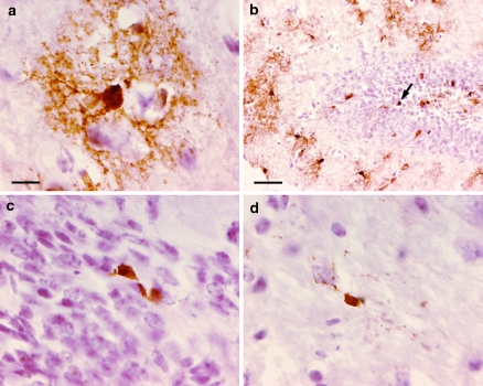
Fig. 4.
Cre-mediated recombination in adult GFAP-CreER TM A; Z/EG brains by immunohistochemistry for GFP. Mice were treated with tamoxifen as in Fig. 3. (a) Cerebral cortex. The stellate appearance of a cortical GFP-stained cell resembling an astrocyte is presented. (b) Dentate gyrus. The arrow indicates a GFP-stained cell located in the inner layer of the dentate gyrus which may represent a neural precursor cell. (c) Subventricular zone. These GFP-positive cells may represent self-renewing B cells of the subventricular zone. (d) Corpus callosum. Scale bar in (a) is 10 μm and applies to panels (a, c, d). Scale bar in (b) is 50 μm

