Fig. 6.
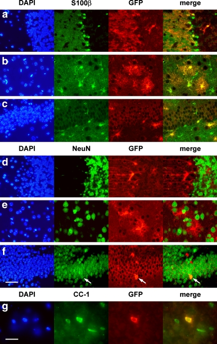
Identification of cells undergoing Cre-mediated recombination in adult GFAP-CreER TM A; Z/EG brains by double immunofluorescence. Mice were treated with tamoxifen as in Fig. 3. (a–c) Staining with an astrocyte marker and anti-GFP. Co-immunodetection of S100β (green) and GFP (red) is demonstrated in the cerebellum (a), cerebral cortex (b) and dentate gyrus (c). (d–f) Staining with a neuronal marker and anti-GFP. There is no co-immunodetection of NeuN (green) and GFP (red) in cerebellum (d) and cortex (e) while occasional granule neurons of the dentate gyrus (f) express both markers (arrow). (g) Staining with an oligodendrocyte marker and anti-GFP. Cells staining with both CC-1 (green) and GFP (red) were readily identified in the corpus callosum. Scale bar in (f) is 50 μm and applies to panels (a–f). Scale bar in (g) is 20 μm. DAPI: 4′,6-diamino-2-phenylindole (blue). Immunophenotyping results were identical in the GFAP-CreER TM B line (data not shown)

