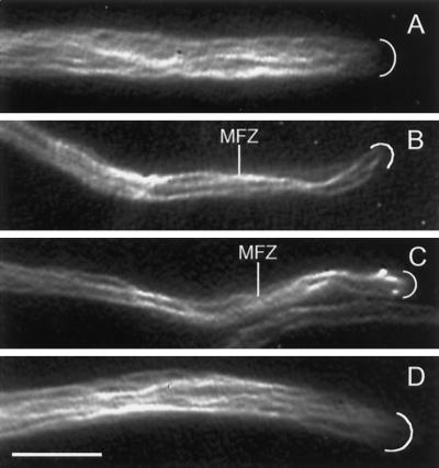
Figure 5.
Fluorescence videomicrographs showing immunocytochemical localization of MTs in hyphal tip cells of the wild type (A), the NhKIN1− deletion mutant TSN25 (B and C), and the NhKIN1+ transformant TSN20 (D). The MT distribution in the mutant cells appears normal, with MTs present throughout the mitochondrion-free zone (MFZ). White arcs mark the positions of the hyphal apices. Bar, 5 μm.

