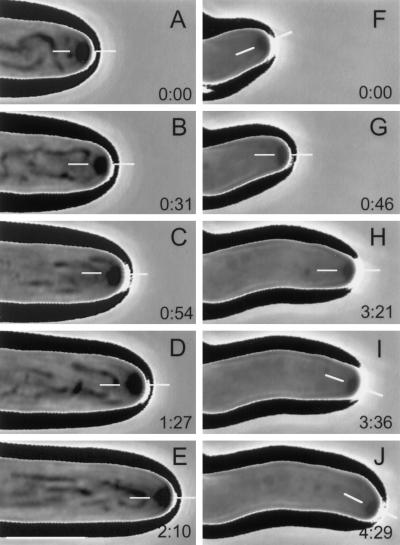
Figure 6.
Phase-contrast, time-lapse videomicrographs of growing hyphal tip cells illustrating the effects of kinesin deletion on positioning of the Spitzenkörper and, consequently, on hyphal morphogenesis. Elapsed time (min:sec) is shown in the lower right corner of each panel, and thin, white bars in each panel indicate the position of the Spitzenkörper relative to the hyphal apex. (A–E) A representative tip cell of the ectopic transformant-control. The central position of the Spitzenkörper was maintained throughout, resulting in a straight hypha. (F–J) A representative tip cell of the kinesin mutant. Central positioning of the Spitzenkörper produced a short, straight segment of hyphae (F). Then the position of the Spitzenkörper was shifted (G) and maintained long enough to produce another straight segment of hypha (H) with a different growth orientation. Finally, another shift of the Spitzenkörper (I) produced another short segment of hypha (J) with yet another growth orientation. Bar (in the lower left corner of panel E), 5 μm.

