
Figure 1.
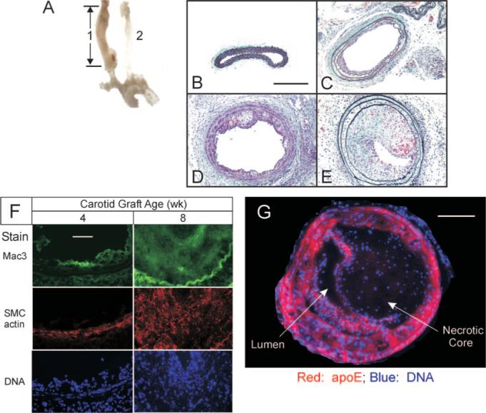
Atherosclerosis progresses rapidly in congenic carotid grafts implanted in apoe−/− mice. A, The 8-mm carotid interposition graft (1), thickened by atherosclerosis, was harvested 8 weeks postoperatively, along with the aortic arch and contralateral carotid (2). Cross-sections of carotid grafts are shown (original magnification ×220) from specimens harvested 0 (B), 4 (C), 6 (D), and 8 (E) weeks postoperatively, and stained with a modified connective tissue stain. Scale bar=200 μm. F, Carotid grafts harvested 4 or 8 weeks postoperatively were frozen, and serial sections were immunostained for either macrophages (Mac3) or SMC α-actin, and counterstained with Hoechst 33342 (DNA). The luminal surfaces are oriented upward in each panel. Images of single specimens are representative of 3 grafts of each postoperative age. Scale bar=50 μm (original magnification ×440). G, The 8-week-old carotid grafts were stained for DNA and apoE, the staining pattern for which corresponds to SMC actin and SMC myosin heavy chain (supplemental Figure IIA and data not shown, n=3). Scale bar=100 μm.