
Figure 3.
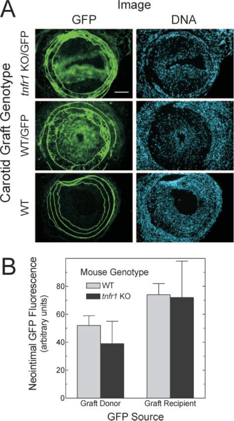
Arterial wall expression of TNFR1 does not affect the prevalence of atherosclerotic lesion cells derived from the arterial wall or circulating progenitors. Carotid grafting was performed as in Figure 2, except that either donor or recipient mice were transgenic for ubiquitous GFP expression (/GFP). A, Frozen sections of grafts were stained with Hoechst 33342 and imaged sequentially for endogenous GFP fluorescence (green) and DNA fluorescence. Images are representative of ≥4 grafts of each type (original magnification ×220); scale bar=100 μm. B, Neointimal green fluorescence in non-GFP specimens (nonspecific) was subtracted from that in GFP specimens (total fluorescence) to obtain specific GFP fluorescence. This value was divided by the cognate value for neointimal DNA fluorescence to obtain GFP/DNA (arbitrary units). These neointimal GFP/DNA values were averaged among grafts of each indicated type, and the mean±SE of ≥4 independent graft specimens is presented.