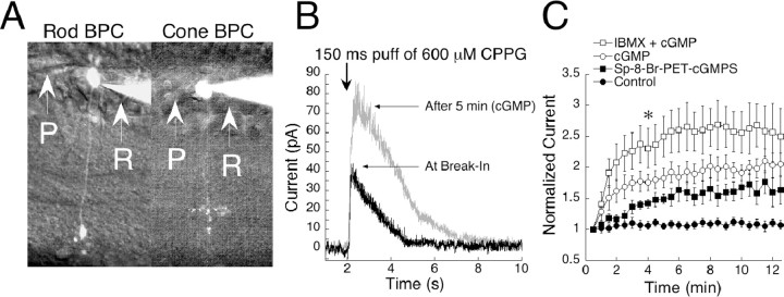
Figure 1.
cGMP potentiates On bipolar cell responses in mouse. A, Light microscope images of a rod bipolar cell (left) and a cone On bipolar cell (right) filled with Alexa Fluor 488 and visualized with fluorescence. The two cell types can be distinguished by the position of their terminals in the inner plexiform layer. P, Puffer pipette; R, Recording pipette. B, Raw traces from a cell filled with 1 mm cGMP and stimulated with a 150 msec sim-flash at the 2 sec mark. As cGMP diffused into the cell, the amplitude was potentiated by approximately twofold. C, During 15 min of recording, cells dialyzed with cGMP potentiated by 180% (n = 12). Responses of control cells remained stable in amplitude throughout the session (n = 11). When IBMX was included in the pipette together with cGMP (n = 7), responses potentiated more than with cGMP alone. Inclusion in the pipette of the non-hydrolyzable cGMP analog sp-8-Br-PET-cGMPS (n = 3) caused an amplification of 60%. Data from each condition were individually normalized to the initial response, and the results were averaged. The plot represents the mean ± SEM for each time point. *p < 0.001, significance at 4 min for all conditions compared with baseline.

