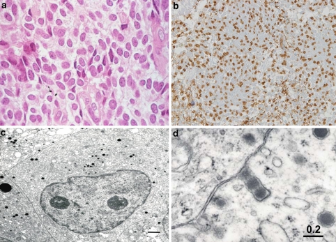
Fig. 7.
PRL producing (cell) adenoma (PRLoma). The tumor cells of hematoxylin eosin (H&E) staining (a) and IHC of PRL, which shows condense in the Golgi regions near the nuclei (b). In electron microscopy, prominent Golgi saccules and a few small NSGs are seen (c). d shows the extrusion of secretory granules, so called “misplaces exocytosis”

