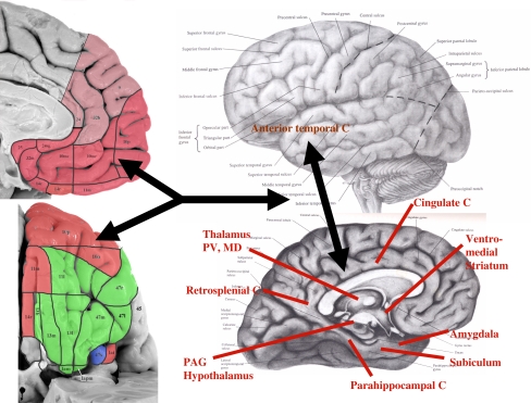
Fig. 1.
Regions and anatomical projections that form the extended visceromotor network. The cytoarchitectonic subdivisions of the human orbital (upper left) and medial prefrontal cortical surfaces (lower left) are distinguished here as being predominantly in the visceromotor (pink) or sensory (green) networks described in (Ongür and Price 2000). These portions of the figure are modified from Ongür et al. (2003), with the lighter shade of pink reflecting more recent work regarding the portions of the medial wall that share the connectional features of the visceromotor network. The area shown in blue, the sulcal portion of BA 47 [47 s; which corresponds to orbital portion of Walker area 12 (i.e., 12o) of the monkey; see Fig. 2], shares features of both the visceromotor and sensory networks. This region and the anterior (agranular) insula (Ia) continue into the lateral cortical wall, so are better viewed in the coronal sections shown in Ongür et al. 2003). The major structures that receive efferent projections from the visceromotor component of the OMPFC are indicated on the right panel over the brain diagram. These include the posterior cingulate cortex, the anterior temporal cortex, and the entorhinal and parahippocampal cortex, all of which are implicated in the “default system” (Hsu and Price 2007; Kondo et al. 2003, 2005; Price 2007; Saleem et al. 2007, 2008). C cortex, MD mediodorsal nucleus of the thalamus, PAG periaqueductal gray; PV periventricular nucleus of the thalamus

