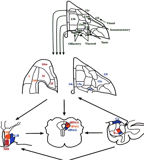
Fig. 2.
Architectonic maps of the orbital (upper and right-center panels) and medial prefrontal cortical surfaces (left-center panel) of the macaque brain, modified from Carmichael and Price (1994). The upper panel shows the areas hypothesized to form the “sensory” network of the orbital cortex based upon their afferent connections with various sensory domains, which are indicated next to each set of regions. This sensory network projects into the “visceromotor” network (middle panel). This latter network shares extensive, reciprocal connections with the amygdala, periaqueductal gray and hypothalamus (shown in coronal sections at the lower right, center and left, respectively), areas which play major roles in organizing or mediating the endocrine, autonomic, and behavioral aspects of emotional behavior. The specific cytoarchitectonic areas of the visceromotor component of the orbitomedial PFC are color coded according to the specific nuclei of the amygdala and hypothalamus or the column of the PAG to which they predominantly project (Carmichael and Price 1995; Ongür and Price 1998; Floyd et al. 2000, 2001). Bvl ventrolateral part of the basal nucleus of the amygdala, Ce central nucleus of the amygdala, DH dorsal hypothalamic area, LH lateral hypothalamic area, MH medial hypothalamic area; dlPAG, lPAG, vlPAG dorsolateral, lateral, and ventrolateral columns of the PAG, respectively

