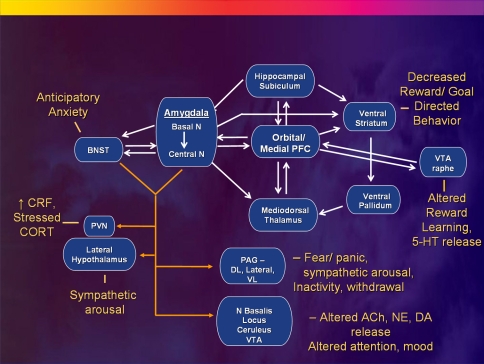
Fig. 5.
Anatomical circuits involving the medial PFC (MPFC) and amygdala reviewed within the context of a model in which MPFC dysfunction results in disinhibition of limbic transmission through the amygdala, yielding the emotional, cognitive, endocrine, autonomic and neurochemical manifestations of depression. The basolateral amygdala sends efferent projections to the central nucleus of the amygdala (ACe) and the bed nucleus of the stria terminalis (BNST). The efferent projections from these structures to the hypothalamus, periaqueductal gray (PAG), nucleus basalis, locus ceruleus, raphe and other diencencephalic and brainstem nuclei then organize the neuroendocrine, neurotransmitter, autonomic, and behavioral responses to stressors and emotional stimuli (Davis and Shi 1999; LeDoux 2003). The MPFC shares reciprocal projections with all of these structures (although only the connections with the amygdala are illustrated) which function to modulate each component of emotional expression (Ongür et al. 2003). Impaired MPFC function thus may disinhibit or dysregulate limbic outflow through the ACe and BNST. Solid white lines indicate some of the major anatomical connections between structures, with closed arrowheads indicating the direction of projecting axons. Solid yellow lines show efferent pathways of the ACe and BNST, which generally are monosynaptic, but in some cases are bisynaptic connections (e.g., Herman and Cullinan 1997). Other abbreviations: 5-HT serotonin, ACh acetylcholine, DA dopamine, DL dorsolateral column of PAG; N nucleus, NE norepinephrine, NTS nucleus tractus solitarius, PVN paraventricular N of the hypothalamus, VL ventrolateral column of PAG, VTA ventral tegmental area. Reproduced from Drevets (2007)

