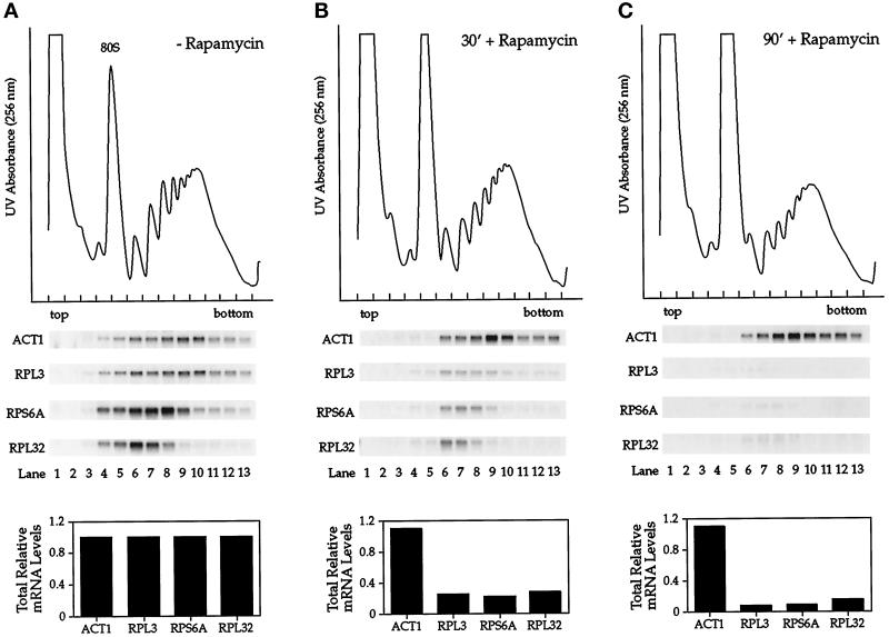
Figure 1.
The fate of r-protein mRNAs in polyribosomes after treatment of yeast cells with rapamycin. Exponentially growing cells were treated with drug vehicle alone for 90 min (A) or with rapamycin for either 30 min (B) or 90 min (C). Cell extracts were prepared, and polyribosomes were separated by sucrose density ultracentrifugation followed by fractionation. Top, a UV absorbance profile was recorded by scanning at 256 nm to display the polyribosome profile. Middle, RNA was isolated from individual fractions and analyzed by Northern blotting, probing for the specified mRNAs. Bottom, the total relative amount of each mRNA present in the different cell extracts is indicated, normalized to levels present in cells treated with drug vehicle alone.

