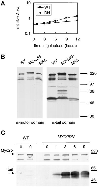
Figure 2.
Overexpression of the tail of Myo2p causes a dominant–negative phenotype. MYO2DN (DN) and wild-type (WT) cells were grown in YP-glycerol and then shifted to YP-galactose for varying amounts of time. At each time point the A600 was measured (A), and protein samples were made (C). (A) The growth of cells transformed with the GAL1 vector (WT) or GAL1–Myo2p tail (MYO2DN) was monitored by reading the A600 at 1, 3, 4.5, 6, and 9 h after a shift to galactose-containing medium. (B) Both motor- and tail-domain antibodies are specific for Myo2p. Wild-type (WT), Myo2-GFP (M2-GFP), and Myo4 knock-out (M4Δ) cells were lysed, and gel samples from each strain were separated by SDS-PAGE and immunoblotted using affinity- purified anti-Myo2p motor domain antibody or anti-Myo2p tail antibody. The immunoblot using the Myo2p-tail antibody was overexposed so that the ∼70 kDa GFP-specific breakdown product could be visualized. Molecular weight markers are indicated on the right-hand side of the immunoblots. (C) Immunoblots using anti-Myo2p head or an anti-HA antibody reveal expression levels of the endogenous Myo2p and tail, respectively, at each time point. Equal amounts of total protein were loaded in each lane. Relative amounts of overexpressed tail were determined using scanning densitometry. Molecular weight markers are indicated on the right-hand side of the immunoblots.

