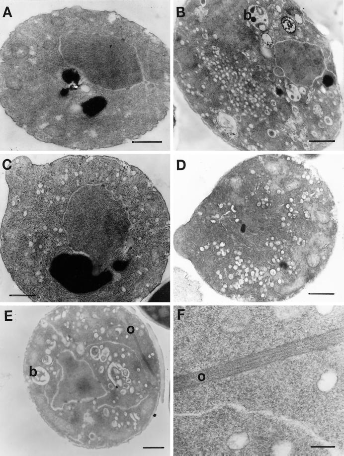
Figure 4.
Thin-section EM of MYO2DN. Wild-type (WT) cells were grown in YP-galactose for 6 h (A). myo2–66 cells were grown in YP-dextrose at 37°C for 20 min (B). MYO2DN cells were grown in galactose for 1 (C), 3 (D), or 6 h (E and F). Berkeley bodies (b) were observed in myo2–66 after 20 min at the restrictive temperature and MYO2DN cells at the 6-h time point. Ordered arrays (o), which may represent actin bars, were observed in MYO2DN at the 6-h time point (E and F). Scale bar, 500 nm (A–E); 100 nm (F).

