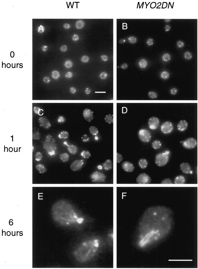
Figure 5.
Actin localization in MYO2DN. Wild-type (WT) and MYO2DN cells were grown in glycerol (A and B) and shifted to galactose for 1 (C and D) or 6 h (E and F), fixed, and stained with anti-actin antibody. After a 1-h shift to galactose, actin patches were polarized to the buds of dividing cells, and actin filaments could be detected. After 6 h of growth in galactose, 85% (n = 107) of the cells observed had abnormal actin bars or large patches. Scale bar, 5 μm (A–D); 5 μm (E and F).

