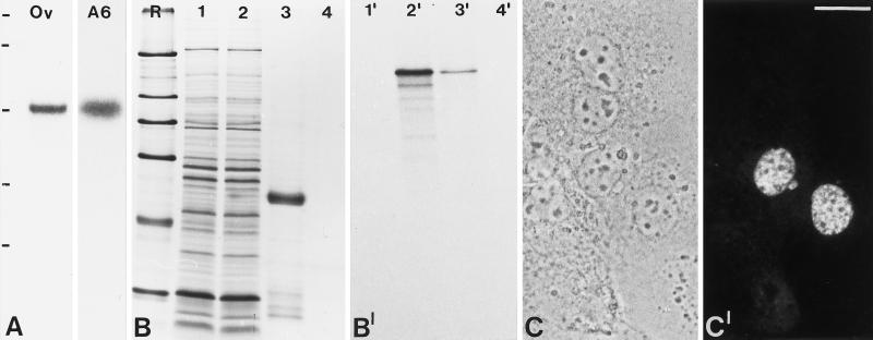
Figure 2.
Molecular characterization of the cDNA clone encoding the 146-kDa protein. (A) Identification of specific mRNAs by Northern blot analysis. Poly(A)+-RNA from Xenopus oocytes (Ov) and Xenopus A6 cells (A6) was separated in an agarose gel, transferred to a membrane, and probed with antisense cRNA derived from clone pBT B2-Xen. Note the specific reaction with a ∼4.4 kb RNA. RNA-size markers of 7.5, 4.4, 2.4, and 1.4 kb are indicated on the left (from top to bottom). (B) Coomassie blue staining of SDS-PAGE–separated rabbit reticulocyte lysates after in vitro transcription/translation in the absence (lane 1) or presence of the pBT B2-Xen template (lane 2). Lanes 3 and 4 show immunoprecipitates of translated protein in the presence or absence of antibody B2.4–1, respectively. R, reference proteins: 205, 116, 97.4, 66, 45, and 29 kDa (from top to bottom). (B′) Corresponding autoradiograph of translation products and immunoprecipitates. (C) Phase-contrast microscopy of cultured human hepatocellular carcinoma (PLC) cells used to determine the subcellular localization of the 146-kDa Xenopus protein carrying an amino-terminal myc tag in transfection experiments. (C′) Corresponding immunofluorescence using mAb 9E10 recognizing the myc tag. Bar, 20 μm

