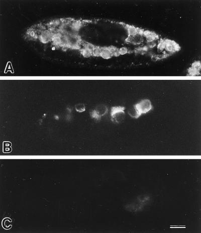
Figure 8.
Immunofluorescence in cells labeled with mAbs against C5 and L1 antigens. (A) Cell fixed, acetone-permeabilized, and then labeled with mAb against C5 antigen. (B) The cell was briefly and weakly permeabilized with Triton X-100 first and then incubated with mAb against C5 antigen before fixation and secondary antibody incubation. The positive result shows that the mAb was able to bind to the antigenic sites on the cytosolic surfaces of the DVs. (C) This cell was similarly treated as in B but was exposed to mAb against L1, an antigen known to be on the luminal side of the DV-II membranes. The negative result shows that this mAb was not able to penetrate the DV-II membrane to get to the antigenic site on the luminal side of the DV membranes. Bar, 20 μm.

