
Figure 4. Efflux of filamentous UPEC from a bladder superficial epithelial cell.
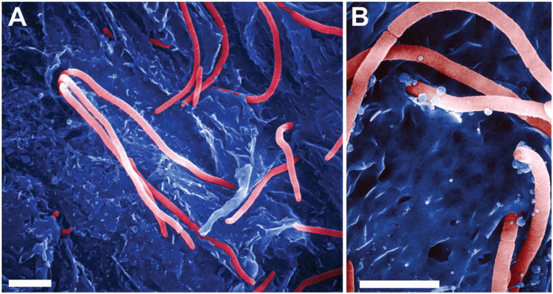
(A and B) Scanning electron microscopy shows filamentous UPEC (a cystitis isolate designated UTI89) emerging from and binding to mouse bladder superficial cells. Scale bars, (A) 5 μm and (B) 3 μm.