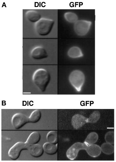
Figure 6.
GFP–Yck2p becomes concentrated at shmoo tips in pheromone-treated cells and is localized to the site of fusion in mating cells. (A) Cells of strain LRB854 (MATa yck1-Δ1::ura3 GFP:YCK2) were grown to 2 × 10⁷ cells/ml in synthetic medium at 30°C and exposed to alpha mating pheromone in the same medium at a final concentration of 6 μg/ml for 2 h. Cells were observed as described in the legend to Figure 2. (B) GFP:YCK2 haploid cells of strains LRB854 and LRB855 were mixed on solid rich medium and incubated for 3 h at 30°C. Cells were then suspended in synthetic medium and placed on slides for observation by DIC and fluorescence optics. Bar, 2 μm.

