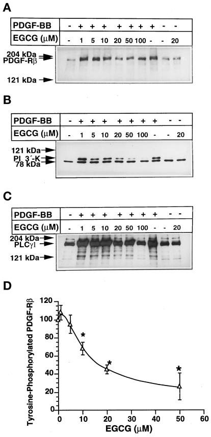
Figure 3.
Effect of PDGF-BB on tyrosine phosphorylation of PDGF-Rβ, PI 3′-K, and PLC-γ1 in EGCG-treated VSMCs. Confluent cells in 75-cm2 flasks were preincubated in serum-free medium in the presence and absence of different concentrations of EGCG. Then the medium was replaced with serum-free medium without EGCG, and VSMCs were stimulated with 50 ng/ml PDGF-BB for 5 min. Then the cells were lysed, and tyrosine-phosphorylated proteins were immunoprecipitated using an anti-phosphotyrosine antibody coupled to Sepharose. Proteins (5 μg) were analyzed by 7.5% SDS-PAGE. Tyrosine-phosphorylated PDGF-Rβ (A), PI 3′-K (B), and PLC-γ1 (C) were detected on the same blot by the enhanced chemiluminescence method using the respective monoclonal antibodies. (D) Laser densitometric analysis of the band densities obtained by three separate experiments showing the effect of PDGF-BB on tyrosine phosphorylation of PDGF-Rβ in VSMCs after treatment with various concentrations of EGCG.

