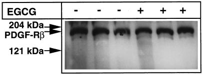
Figure 6.
Effect of EGCG on the PDGF-Rβ amount in VSMCs. VSMCs were cultured in dishes (diameter: 3 cm) and cultivated until confluence. Then the medium was replaced by serum-free medium, and VSMCs were incubated in the presence and absence of 50 μM EGCG for 24 h. VSMCs were then lysed, and 20 μg of protein were analyzed with SDS-PAGE. PDGF-Rβ was detected by enhanced chemiluminescence Western blotting using anti–PDGF-Rβ antibodies.

