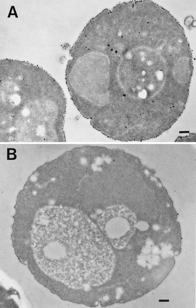
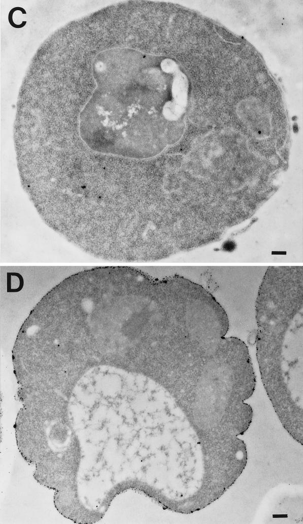
Figure 1.
Positively charged Nanogold can be used as an endocytic marker. Wild-type and end3 mutant strains were grown overnight, converted to spheroplasts, and incubated under various conditions on ice for 15 min followed by a 30-min incubation at RT. The samples were fixed and embedded and thin sections were cut. (A) Sections from wild-type cells incubated with positively charged Nanogold were enhanced with HQ Silver. (B) Sections from end3 mutant cells incubated with positively charged Nanogold and enhanced. (C) Sections from wild-type cells incubated in the absence of positively charged Nanogold, but enhanced. (D) Sections from wild-type cells incubated with positively charged Nanogold in the presence of NaN3 and NaF and enhanced. The cells were visualized in the electron microscope as described in MATERIALS AND METHODS. Bar, 200 nm.


