Figure 3.
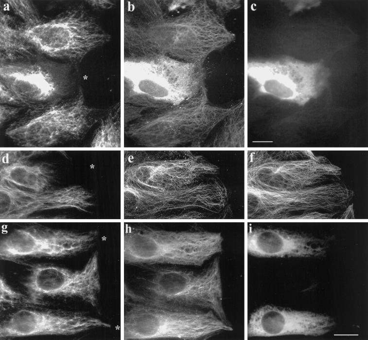
Microinjection of nonpolymerizable, IAA-Glu tubulin causes collapse of the IF network. 3T3 cells at the edge of an in vitro wound were injected with IAA-treated Glu tubulin (from calf brain) at 140 μM (a–c) or at 70 μM (d–f) or with untreated Glu tubulin at 100 μM (g–i). A marker protein, human IgG (2 mg/ml), was coinjected in all cases. Cells were fixed 2 h after injection and immunofluorescently stained to reveal vimentin (a, d, and g), Glu tubulin (b, e, and h), human IgG (c and i), and Tyr tubulin (f). The asterisks in a, d, and g indicate the cell periphery of the injected cell (the human IgG marker is not shown for d–f). Note that in the cells injected with IAA-Glu the IFs are collapsed around the nucleus and do not extend to the cell edge, whereas in cells injected with untreated, assembly-competent Glu tubulin the IFs remain extended in the cytoplasm. Microinjection of human IgG alone had no apparent effect on the distributions of either the MTs or the IFs (our unpublished results). Bar, 10 μm.

