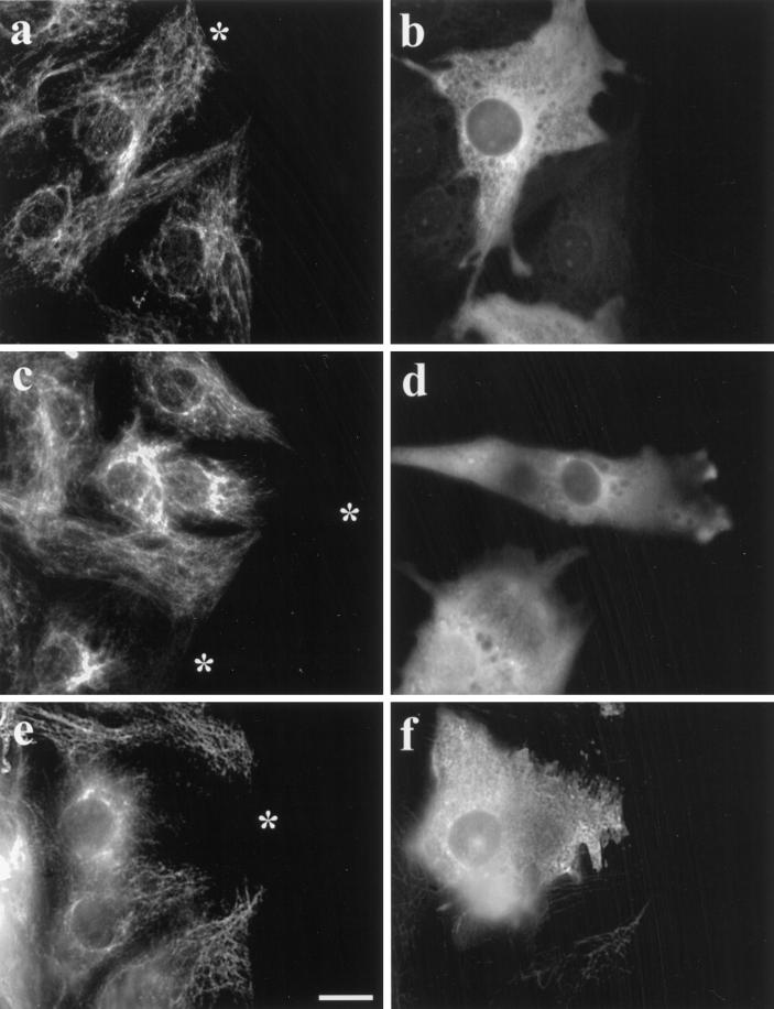
Figure 6.
C-terminal fragments of α-tubulin induce collapse of IFs. Wound-edge cells were microinjected with N-terminal (α-N) or C-terminal (α-C and α-C Glu) α-tubulin trypsin fragments, incubated for 2 h at 37°C, fixed, and immunofluorescently stained for vimentin IFs (a, c, and e), human IgG marker (b and d), Glu tubulin (f), and Tyr tubulin. IFs are unaltered in cells injected with 70 μM α-N (a and b) but are collapsed to a perinuclear region in cells injected with 145 μM α-C (c and d) or 235 μM α-C Glu (e and f). Asterisks denote the peripheral edge of injected cells. Bar, 10 μm.

