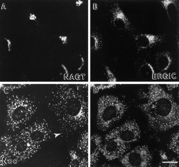
Figure 10.
Effect of nocodazole treatment on the distribution of NAGT-I and ERGIC-53. Vero cells treated with nocodazole for 0 min (A and B) or 4 h (C and D) were processed for indirect immunofluorescence using polyclonal anti-myc antibodies to localize NAGT-I-myc (rhodamine channel, A and C) and the monoclonal anti-ERGIC-53 antibody G1/93 (FITC channel, B and D). White arrowhead in C points to an example of NAGT-I-positive cytoplasmic patch which appeared negative for ERGIC-53. Bar, 20 μm.

