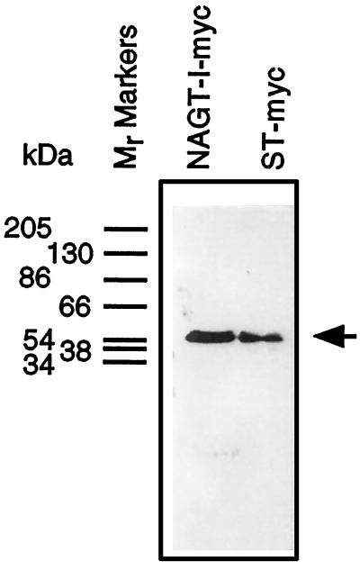
Figure 4.
Electrophoretic characterization of NAGT-I and ST-myc expressed in stably transfected Vero cells. Golgi fractions were prepared from NAGT-I- and ST-myc Vero cells and solubilized, and equal aliquots of the fractions were applied to wells of a SDS-polyacrylamide gel. After transfer to nitrocellulose, filters were probed with 9E10 mAb diluted in blocking buffer and the primary antibody was then localized by ECL using HRP-conjugated secondary antibody. The position of molecular weight markers are indicated on left side. The prestained mobility standards are: α2-macroglobulin, 205 kDa; β-galactosidase from Escherichia coli, 130 kDa; fructose-6-phosphatase kinase from rabbit muscle, 86.5 kDa; pyruvate kinase from chicken muscle, 66 kDa; fumarase from procine heart, 54 kDa; lactate dehydrogenase from rabbit muscle, 38 kDa; and triosephosphatase from rabbit muscle, 34.2 kDa.

