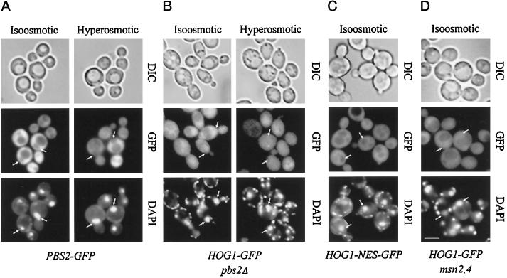
Figure 4.
The MAPKK Pbs2 is not required for anchoring or basal nucleocytoplasmic cycling of Hog1. (A) Pbs2 appears constitutively localized in the cytoplasm. Strain VRY 10 (pbs2Δ) was transformed with pVR28 (2 μm PBS2–GFP), and transformants were grown in appropriate selective medium (iso-osmotic); where indicated, hyperosmotic stress was applied (0.4 M NaCl, 5 min; hyperosmotic). The positions of nuclei are indicated by arrows. (B) Strain VRY 10 (pbs2Δ) was transformed with pVR65-WT (HOG1–GFP). Cells with nuclear staining were counted after growing the strain to logarithmic phase in selective medium (iso-osmotic) and subjecting them to hyperosmotic conditions as described above. (C) Hog1–NES–GFP does not show basal nuclear staining under nonstress conditions. Strain K4327 (hog1Δ) was transformed with centromer plasmid pVR65–NES (HOG1–GFP fused to PKI NES), and cellular distribution of Hog1 was determined after growing them to logarithmic phase in appropriate selective medium (iso-osmotic). (D) Hog1 basal cycling is not dependent on Msn2 and Msn4 transcription factors. Strain YM24 (msn2,4Δ) was transformed with centromer plasmid pVR65 (HOG1–GFP), and cellular distribution of Hog1 was determined after growing cells to logarithmic phase in appropriate selective medium (iso-osmotic). Bar, 5 μm.

