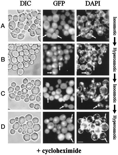
Figure 7.
Cellular distribution of Hog1 during stress and adaptation is independent of de novo protein synthesis. Logarithmically growing strain K4327 (hog1Δ) transformed with centromer plasmid pVR65–WT (HOG1–GFP) was preincubated with cycloheximide (0.1 mg/ml) for 2 h before microscopy. Pictures were taken from cells incubated under iso-osmotic (selective medium) (A) or hyperosmotic conditions (5 min after addition of NaCl to a final concentration of 0.4 M) (B). (C) Cells from B were left to adapt for 60 min and then reexposed to hyperosmotic stress (5 min, final concentration of NaCl 0.8 M) (D). The positions of nuclei are indicated by arrows. Bar, 5 μm.

