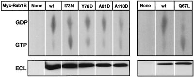
Figure 4.
Guanine nucleotide composition of Myc-Rab1B constructs expressed in HEK-293 cells. In two separate experiments (left and right panels), 293 cells were labeled for 5 h with [32P]orthophosphate, beginning 24 h after transfection with the indicated Myc-Rab1B constructs (see MATERIALS AND METHODS). Myc-tagged proteins were immunoprecipitated and 50% of each sample was subjected to immunoblot analysis with antibody to Rab1B using chemiluminescent detection (ECL, lower panel). 32P-labeled guanine nucleotides in the immunoprecipitates were detected by subjecting 25% of each sample to TLC (upper panel). Radioactivity was quantified by scanning with a phosphorimager and GDP:GTP ratios were determined. The results are summarized in Table 1.

