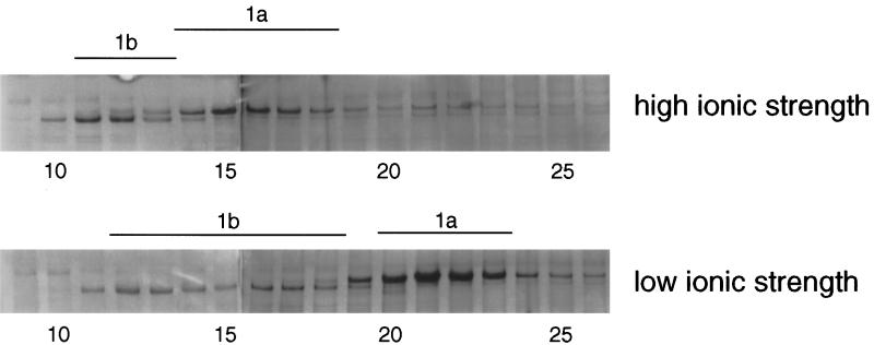
Figure 5.
Effect of ionic strength on the sedimentation patterns of cytoplasmic dyneins. Testis cytoplasmic dynein prepared by ATP extraction of microtubules was sedimented in 5–20% sucrose gradients made in high ionic strength (top) or low ionic strength (bottom). The gels of the samples were stained with silver. Each panel shows the DHC regions of two gels joined together. In high ionic strength, the DHC1a sedimented in a peak centered in fractions 15–16, and the anti-1b-immunoreactive protein sedimented in a tight peak centered in fractions 11–12. In low ionic strength, the DHC1a sedimented faster in the gradient, centered in fractions 21–22, and the anti-1b immunoreactive protein sedimented as a broad peak in fractions 11–18.

