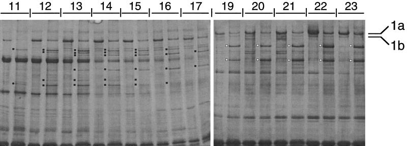
Figure 7.
V1 photolysis of cytoplasmic dynein heavy chains separated in a sucrose gradient. Testis cytoplasmic dynein prepared by ATP extraction of microtubules was sedimented through a 5–20% sucrose gradient made in low ionic strength. One hundred-microliter aliquots of individual gradient fractions 11–17 and 19–23 were subjected to V1 photolysis. Equal volumes of untreated and V1-photolyzed samples were resolved in 6.7% SDS-polyacrylamide gels and stained with silver. Two gels were joined together. The principal V1-photolytic products are marked with dots. The 1b-like fractions (11–17) yielded six V1-photolytic products (filled dots). The pattern of products changed through the gradient. DHC1a (fractions 19–23) yielded two V1-photolytic products (open dots).

