Figure 2.
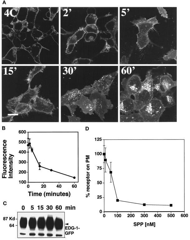
Exogenous SPP-induced internalization of EDG-1–GFP. (A) Confocal fluorescence microscopy. HEK293 cells stably expressing the EDG-1–GFP polypeptide were preincubated in CFBS for 2 d and treated with 100 nM SPP at 4°C for 15 min (4C). Cells were then shifted to 37°C for various time periods (2–60 min), fixed, and imaged in a confocal fluorescence microscope. Bar, 10 μm. (B) Quantitative analysis of EDG-1–GFP localization on the plasma membrane. Digitized fluorescence intensity on the plasma membrane after SPP treatment was quantitated as described and analyzed from six randomly selected fields. (C) Immunoblot analysis. HEK293 cells stably expressing the EDG-1–GFP polypeptide were starved in CFBS for 2 d and stimulated with 100 nM SPP for various times, cell extracts were prepared, and immunoblot analysis with the anti-GFP antibody was conducted as described. (D) Dose–response of SPP-induced internalization of EDG-1–GFP. HEK293 cells stably expressing the EDG-1–GFP polypeptide were starved in CFBS for 2 d, treated with the indicated concentrations of SPP at 37°C for 30 min, fixed, and imaged in a confocal fluorescence microscope. Digitized fluorescence intensity on the plasma membrane after SPP treatment was quantitated as described and analyzed from six randomly selected fields. Percent fluorescence intensity on the plasma membrane (PM) is shown.

