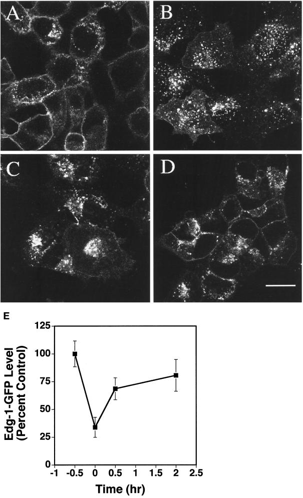
Figure 4.
Recycling of the EDG-1–GFP polypeptide. HEK293 cells expressing EDG-1–GFP polypeptide were subjected to various treatments and imaged by confocal microscopy. (A) Cells were treated with cycloheximide (CHX) for 30 min to block protein synthesis. (B) Cells were then stimulated with 100 nM SPP for 30 min in the presence of CHX. Note the accumulation of EDG-1–GFP in intracellular vesicles. (C and D) Exogenous SPP was washed out, and media containing CHX was replaced and incubated for 30 min (C) and 120 min (D) at 37°C. Note that the EDG-1–GFP polypeptide recycles back to the plasma membrane at 120 min. Bars, 10 μm. (E) Quantitative analysis of internalization and recycling of EDG-1–GFP is shown. Digitized fluorescence intensity from the plasma membrane was quantitated by the imaging software and normalized to the prestimulation level. Data represent mean ± SD of fluorescence intensities of six randomly selected fields.

