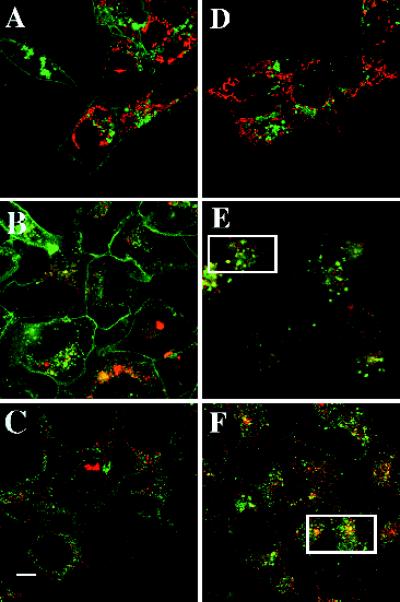
Figure 5.
Colocalization of intracellular organelles and EDG-1–GFP. HEK293 cells expressing EDG-1–GFP were grown in 10% FBS, incubated with various organelle-specific dyes in the presence or absence of SPP (100 nM) for 1 h, and imaged by confocal microscopy to localize the EDG-1–GFP polypeptide (488 nm) and the organelle-specific dyes (568 nm). (A and D) Untreated (A) and SPP-treated (D) cells were incubated with TMRE dye (50 nM) for 15 min at 37°C to label the mitochondria. (B and E) Untreated (B) and SPP-treated (E) cells were labeled with Lysotracker dye to label the lysosomal structures. (C and F) Untreated (C) and SPP-treated (F) cells were preincubated for 0.5 h with Texas red–labeled transferrin to visualize endosomal structures. Rectangles in E and F indicate cells in which significant colocalization is observed. Bar, 10 μm.

