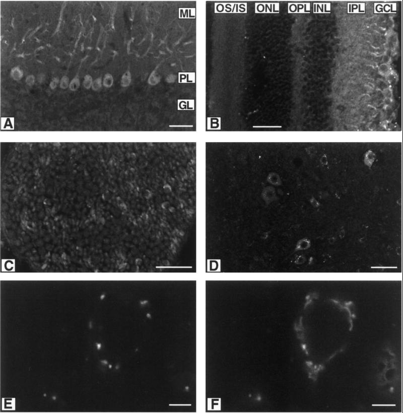
Figure 3.
Cellular localization of the KIF3C motor by immunofluorescence microscopy. Anti-KIF3C stain: (A) section of cerebellum, (B) section of retina, (C) cross section of sciatic nerve, and (D) cross sections of spinal cord. Double staining of cross section of spinal cord with anti-KIF3C (E) and anti-giantin (F). ML: molecular layer, PL: Purkinje cell layer, GL: granular layer; OS/IS; outer and inner segments, ONL: outer nuclear layer, OPL: outer plexiform layer, INL: inner nuclear layer, IPL: inner plexiform layer, GCL: ganglion cell layer. Scale bar: 50 μm in A–D; 5 μm in E and F.

