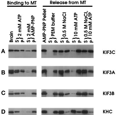
Figure 6.
Microtubule sedimentation analysis of KIF3C. KIF3C binding to microtubules (left panels): soluble protein from total brain extract was incubated with GTP and Taxol to boost microtubule polymerization. After microtubule assembly, extracts were supplemented with ATP or AMP-PNP. KIF3C release from microtubules (right panels): the microtubule pellet (AMP-PNP pellet) was extracted with PEM buffer with or without 0.5 M NaCl, 10 mM ATP, or 0.5 M NaCl plus 10 mM ATP. Pellet (P) and supernatant (S) were separated by centrifugation. An equal percentage of each fraction was analyzed. KIF3C was detected with anti-KIF3C by Western blot analysis (A). The same blots were used for detecting KIF3A (B), KIF3B (C), KHC (D) with anti-KIF3A (Kondo et al., 1994), anti-KIF3B, and a monoclonal antibody Suk4 (Ingold et al., 1994), respectively.

