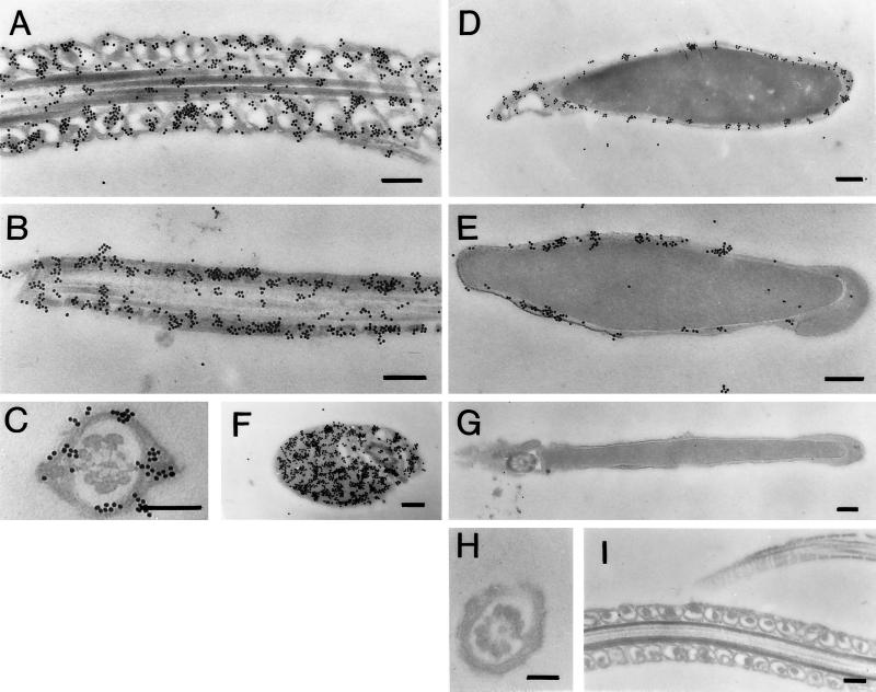
Figure 8.
Immunoelectron microscopic localization of HK1-sc in mouse sperm. Sperm were collected and prepared as described in MATERIALS AND METHODS. Panels A–F are micrographs of sections probed with a 1:200 dilution of purified α-gcs IgG. Panels G–I are micrographs of sections probed with a 1:200 dilution of purified α-gcs IgG preabsorbed with the peptide against which it was made. In all cases, a secondary goat anti-rabbit antibody conjugated to 18-nm colloidal gold particles was used for labeling. (A) A longitudinal section of the midpiece of the flagellum showing labelling associated with the mitochondria. (B) A longitudinal section of the principal piece of the flagellum showing labelling associated with the fibrous sheath. (C) A cross-section of the principal piece of the flagellum showing labelling associated with the fibrous sheath. (D) An oblique section through the posterior head showing labelling in the region of the membranes, which often appeared organized into discrete clusters. (E) A transverse section through the head, including a portion of the equatorial region, showing labelling associated with the membranes of the anterior and posterior sperm head. (F) A cross-section of the midpiece of the flagellum showing labelling of a cytoplasmic droplet. (G) A longitudinal section of a preabsorbed control of the sperm head. (H) A cross-section of a preabsorbed control of the principal piece. (I) Longitudinal sections of a preabsorbed control of the midpiece and fibrous sheath. Bar, 250 nm, in all panels.

