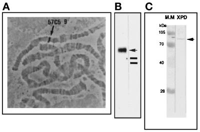
Figure 2.
Panel A shows the DmXPD localization in polytene chromosomes after in situ hybridization. The arrow indicates the position at 57C5–9 in the right arm of the second chromosome. Panel B shows a Northern blot using total RNA from 0- to 24-h embryos using the DmXPD full-length cDNA as a probe. The arrow indicates a band of 3 kb, and the bars indicate the rRNA position in the gel. Panel C shows a Western blot using total protein extracts from embryos of 0–24 h and developed with an anti-DmXPD polyclonal antibody (see MATERIALS AND METHODS). The lane M.M. shows the molecular weight markers; in the DmXPD lane is shown the antibody-detected band from the total embryo extract with the expected DmXPD molecular weight.

