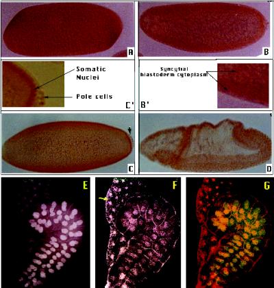
Figure 4.
DmXPD distribution in wild-type embryos and larval tissues. Immunostaining with the anti-DMXPD antibody of embryos in different developmental stages: (A) syncytitial blastoderm; DMXPD is abundant and homogeneously distributed, probably from maternal contribution; (B) cellularization; DMXPD is concentrated in the embryo periphery; (B′), an amplification of panel B; (C) cellular blastoderm; the germ cells are indicated with an arrow, and DmXPD is mostly located in the nuclei of the somatic cells; (C′) an amplificaton of panel C; (D) gastrulation; DmXPD is nuclear in all cells; (E) larval-salivary gland and fat body nuclei stained with cytogreen (which stains DNA); (F) the same preparation as in panel E immunostained with the anti-DmXPD antibody. The arrow indicates cytoplasmic DmXPD in the fat body cells; (G) overlay of panels E and F. Cytogreen-stained DNA appears in green, anti-DmXPD signal is in red, and the overlapping of both signals is in yellow.

