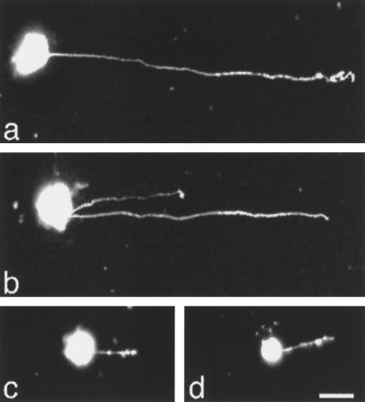
Figure 1.
Epifluorescence micrographs of neurite outgrowth from DRG cells cultured on different L1 fusion protein substrates. DRG cells and neural retinal cells were isolated from E10 chick embryos and labeled with DiI. Cells were cultured for 20 h on coverslips coated with different fusion proteins. DRG cells extended long neurites in response to substrate-coated GST-Ig1-2-3 (a) and GST-Ig4-5-6 (b). When the substrate was precoated with anti-Ig4-5-6 Fab, DRG cells failed to send out long neurites (c). Neural retinal cells cultured on GST-Ig4-5-6 did not extend long neurites (d). Bar, 10 μm.

