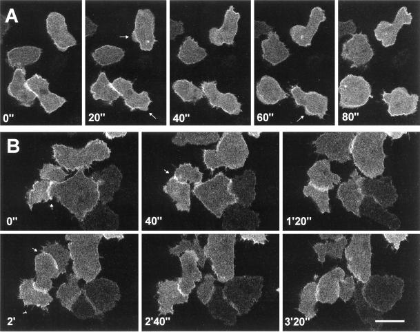
Figure 5.
Localization of GFP–racF1 in vegetative cells. Dictyostelium cells were starved for 3 h and allowed to sit on glass coverslips. Images were taken with a confocal laser scanning microscope. (A) Distribution of GFP–RacF1 during pseudopod formation. Arrows indicate newly forming protrusions. (B) GFP–RacF1 enrichment at sites of cell-to-cell contacts, indicated by arrows. Bar, 10 μm.

