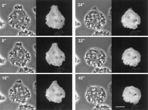
Figure 7.
Dynamics of subcellular localization of GFP–RacF1 during endocytosis. Dictyostelium cells were starved for 3 h and allowed to sit on glass coverslips. Images were taken using a double-view microscope system as described in MATERIALS AND METHODS. Left, phase-contrast image; right, fluorescence image. Note the strong enrichment of GFP–RacF1 at the plasma membrane and the formation of endocytic vesicles. Redistribution of GFP–RacF1 is especially apparent in the vesicle indicated by arrows. Fluorescence around this vesicle is no longer visible after 32 s. Bar, 10 μm.

