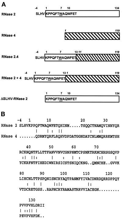
Figure 1.
Structure of hybrid RNases. (A) The RNase 2 portion of a hybrid is depicted as an open rectangle, whereas that of RNase 4 (numbering in italic) is hatched. Trp-7 has been underlined. (B) Comparison of the primary structures of human RNase 2 and porcine RNase 4. Amino acids in common are indicated by a line when they occur at the surface of the protein and by a colon when they are buried.

