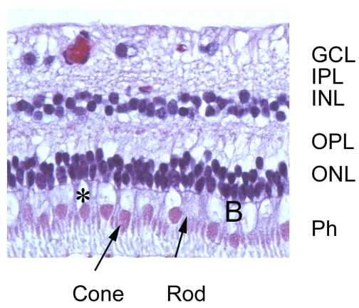
Figure 6.
Retina of a monkey (M. fascicularis) fixed by intraocular injection of 10% neutral buffered formalin. Photographs show cross sections of the retina stained with H&E. Picture shows the retina of an animal treated with DOM (4mg/kg/bw ip). Cell loss and necrosis are present in the INL, GCL and to a lesser extent in the ONL. Vacuoles are easily identified in the Ph cell layer, particularly the cones (*) and in the OPL. Cones and rods in the Ph layer are identified (arrows). There is also marked loss of cell bodies in the GCL. From external to internal: the photoreceptor cell layer (Ph); the outer nuclear layer (ONL); the outer plexiform layer (OPL); inner nuclear layer (INL); inner plexiform layer (IPL); ganglion cell layer (GCL). Objective x40.

