
Figure 3.
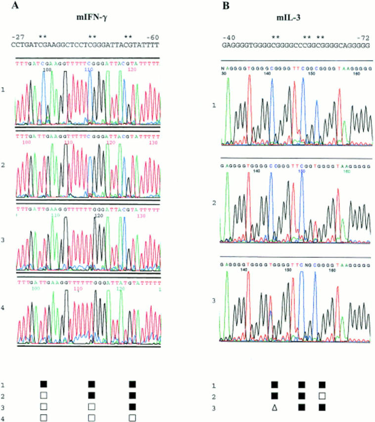
Primary bisulfite genomic DNA sequencing data. Nuclear DNA from clonal cultures of primary mouse CD8+ T cells was purified, bisulfite modified, amplified, and sequenced directly using dye terminator chemistry and automated fluorescent sequence analysis. Sequence results for the noncoding strands of the IFN-γ promoter between bases −27 and −60 in four different clones are shown in the left panel. Sequence results for the noncoding strands of the IL-3 promoter between bases −40 and −72 in three different clones are shown in the right panel. Methylated cytosines are displayed by retained blue cytosine peaks while nonmethylated cytosines are converted by bisulfite modification and PCR to red thymidine peaks. These peaks were scored for each clone and are shown below the chromatograms by the following symbols: ▪, methylated; □, demethylated. The presence of clear coincident cytosine and thymidine peaks, e.g., at position −52 in the third IL-3 sequence, was scored: ▵, partially methylated.