
Figure 2.
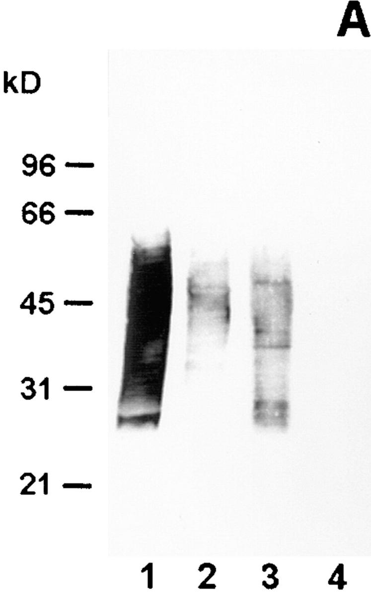
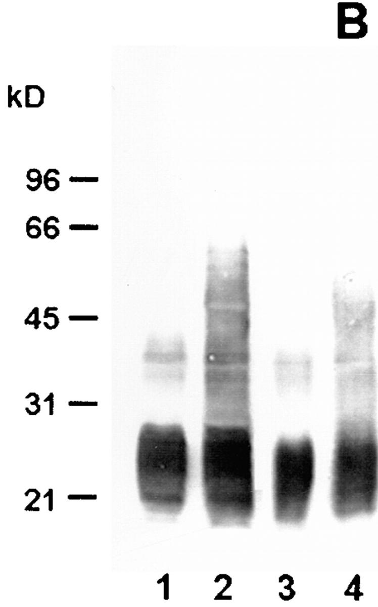
Western blot analysis of wild-type strain RC1 and mutant 811 with mAb 2625 (A) and mAb LPS-1 (B), respectively. Lane 1, wild-type RC1 whole cell lysate; lane 2, mutant 811 whole cell lysate; lane 3, wild-type RC1 2 μg purified LPS; lane 4, mutant 811 2 μg purified LPS. Numbers on the left side indicate molecular masses of a standard protein marker. The molecular mass of L. pneumophila LPS does not correspond to that of the marker proteins, but determination of a relative range of LPS bands was achieved by this method.