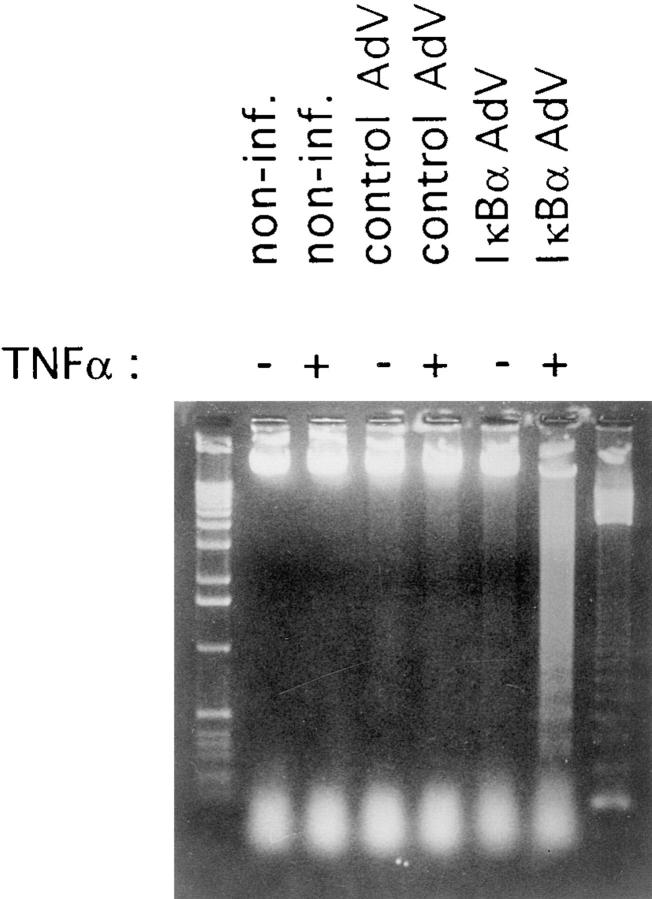
Figure 3.
DNA fragmentation in adenovirus IκBα–infected and TNF-α–stimulated HUVECs. HUVECs were not infected, were infected, with a control adenovirus, or were infected with the recombinant adenovirus-IκBα construct. Noninfected cells and infected cells were left untreated or treated with TNF-α (500 U/ml) for 6 h. Appearance of fragmented genomic DNA was analysed by 1.3% agarose gel electrophoresis. Left lane: 1-kb ladder molecular weight standard; right lane: 123-bp ladder molecular weight standard. Non-inf.: noninfected cells; AdV: adenovirus.

